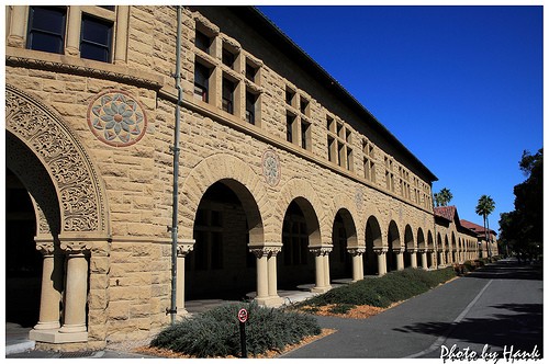
Molecular Imaging Research

- (Stanford University - Hank Ping Han Hsieh)
- Overview
"Molecular Imaging (MI) is a growing biomedical research discipline that enables the visualization, characterization, and quantification of biologic processes taking place at the cellular and subcellular levels within intact living subjects, including patients. MI images depict cellular and molecular pathways and mechanisms of disease present in the context of the living subject. Study of biologic processes in their own physiologically authentic environment is facilitated - MI transcends the requirements for and limitations of in vitro or ex vivo biopsy/cell culture laboratory techniques. It also encompasses 'multiple' image-capture techniques in combination with merging knowledge areas from the fields of cell/molecular biology, chemistry, pharmacology, medical physics, biomathematics, and bioinformatics.
Modern clinical scientist researchers use MI to study the processes of how molecular abnormalities, found in cells, build up to form the basis of disease. This type of study in turn facilitates other important clinical goals of 1) early detection of disease 2) optimizing therapies that aim for certain molecular targets 3) predicting and monitoring response to therapy and 4) monitoring for disease recurrence. Biotechnology companies also use MI to optimize the drug discovery and validation processes." -- [Stanford Medicine]
- Molecular Imaging Research
Molecular imaging differs from traditional imaging in that probes known as biomarkers are used to help image particular targets or pathways. Biomarkers interact chemically with their surroundings and in turn alter the image according to molecular changes occurring within the area of interest. This process is markedly different from previous methods of imaging which primarily imaged differences in qualities such as density or water content. This ability to image fine molecular changes opens up an incredible number of exciting possibilities for medical application, including early detection and treatment of disease and basic pharmaceutical development. Furthermore, molecular imaging allows for quantitative tests, imparting a greater degree of objectivity to the study of these areas. One emerging technology is MALDI molecular imaging based on mass spectrometry.
Many areas of research are being conducted in the field of molecular imaging. Much research is currently centered on detecting what is known as a predisease state or molecular states that occur before typical symptoms of a disease are detected. Other important veins of research are the imaging of gene expression and the development of novel biomarkers. Organizations such as the SNMMI Center for Molecular Imaging Innovation and Translation (CMIIT) have formed to support research in this field. In Europe, other "networks of excellence" such as DiMI (Diagnostics in Molecular Imaging) or EMIL (European Molecular Imaging Laboratories) work on this new science, integrating activities and research in the field. In this way, a European Master Programme "EMMI" is being set up to train a new generation of professionals in molecular imaging.
Recently the term molecular imaging has been applied to a variety of microscopy and nanoscopy techniques including live-cell microscopy, Total Internal Reflection Fluorescence (TIRF)-microscopy, STimulated Emission Depletion (STED)-nanoscopy and Atomic Force Microscopy (AFM) as here images of molecules are the readout.

